
Steffen Baron
Specialist in orthopaedics
Contact Steffen Baron.
At Klinik Manhagen, we treat deformities, joint degeneration, tendon and ligament injuries, sports injuries, nerve entrapment syndromes and fractures of the foot. Our aim is accurate diagnosis and treatment tailored to the individual, whether joint-preserving, stabilising or surgically corrective where appropriate.
Appointment consultation: MVZ Schlump One, Orthopaedics & Trauma Surgery, Schäferkampsallee 18, 20357 Hamburg. Telephone: 040 440606, E-Mail anzeigen.
Surgical assessment consultation: at Klinik Manhagen; arrange an appointment via E-Mail anzeigen (hospital referral required).
Joint-preserving and joint-fusion procedures
Joint-preserving/joint fusion procedures
Reconstructive and corrective procedures
Specialist in orthopaedics
Contact Steffen Baron.
Specialist in orthopaedics and trauma surgery, specialised orthopaedic surgery; Chief Physician
Contact Drs. (NL) Genio Bongaerts.
Specialist in orthopaedics and trauma surgery, knee surgery and sports medicine; Chief Physician
Contact Dr Jan Hennings.

Specialist in orthopaedics and trauma surgery, specialising in hip and knee replacement and robotic surgery
Contact Sebastian Krüsemann.
Specialist in orthopaedics and trauma surgery, Medical Director of Physical Therapy
Contact Dr Sven Gunnar Laabs.
Book an appointment
Specialist in orthopaedics and trauma surgery, manual medicine/chiropractic therapy and emergency medicine; certified foot surgeon
Contact Matthias Roggelin.
Specialist in orthopaedics, emergency medicine and manual medicine/chiropractic therapy
Contact Harald Uphus.
An Achilles tendon spur, also known as a posterior heel spur, is a sign of chronic overloading. The bony spur grows directly into the Achilles tendon and causes pain during physical activity and when wearing tight shoes. Irritation of the Achilles tendon can also cause pain at rest. The tendon is often additionally compressed by a Haglund’s deformity. This lies slightly higher and causes the Achilles tendon to glide over a bony prominence on the heel. Painful inflammation of the bursa can then restrict walking even further.
Physiotherapy may reduce the symptoms, but it rarely relieves the pain completely. This is because the bony spur within the tendon will sooner or later cause pain again. Surgical removal should then be considered. The Achilles tendon has to be released from the heel bone so that the spur can be removed from within the tendon. The bony prominence of the Haglund’s deformity is removed at the same time. The tendon is then reattached to the bone using special anchor systems.
The Achilles tendon takes around eight weeks to grow back into the bone. During this period, patients wear a special boot known as a walker and must use crutches to take weight off the foot for the first four weeks. After eight weeks, patients can generally bear their full weight in a normal shoe. Immobilisation can cause adhesions between the tendon and tendon sheath, so walking may remain restricted for several more weeks. Physiotherapy is often needed during this phase. Sports such as swimming and cycling, and in some cases gym training, are usually possible after around three months; contact sports after around six months.
Achillodynia simply means pain in the Achilles tendon. It can have many different causes. Initially, there may only be functional shortening of the Achilles tendon without any damage to the tendon itself. If symptoms persist, degenerative changes can develop and may become noticeable as thickening of the tendon. These changes are caused by microlesions, or tiny tears, which can be compared with small notches in a porous rubber band as it is stretched. The body attempts to repair these weak areas by forming scar tissue with the help of specific substances in the blood known as growth factors. However, scar tissue is less elastic, so continued loading can cause further lesions and create a vicious circle.
The Achilles tendon has a poor blood supply, meaning that these repair processes take place over many months. As the strongest tendon in the body, it is exposed to substantial loads even during ordinary walking, and the body is sometimes unable to keep pace with the required repairs. This can result in persistent pain during sport and, in some cases, even while walking in everyday life.
Alongside specific exercises for the foot and physiotherapy, extracorporeal shock wave therapy (ESWT) and autologous blood treatment using autologous conditioned plasma (ACP) have proved effective. The two methods promote healing in different ways. Shock wave therapy uses high-energy sound waves that are transmitted from outside the body to the Achilles tendon through an applicator with a gel pad. This stimulates the formation of growth factors and improves blood flow. ACP treatment pursues the same aim of healing but uses a different principle. Blood is taken from the patient and immediately spun at high speed in a centrifuge. The resulting portion of blood contains a high concentration of platelets and their growth factors and is injected directly into the tendon. Both procedures support regeneration of the tendon and generally shorten the healing time. Studies confirm the effectiveness of this approach.*
Non-surgical treatment generally consists of an exercise programme using eccentric training and autologous blood therapy, with shock wave therapy added where appropriate.
*Deans et al., 2012: A prospective series of patients with chronic Achilles tendinopathy treated with autologous-conditioned plasma injections combined with exercise and therapeutic ultrasonography.
Ankle ligament injuries are among the most common sports injuries. They occur when the foot twists during running, particularly on uneven ground or during rapid changes of direction. Many sports are affected: a basketball or volleyball player may land on another player’s foot after jumping, a footballer may lose balance while dribbling, or a runner may stumble on uneven ground such as a forest path with tree roots.
A medical examination is needed to distinguish a sprain from a torn ligament or even an ankle fracture. An X-ray is generally taken and the stability of the ligaments tested during the examination. An MRI may also be needed if the findings are not clear.
Repeated ligament injuries, or ligaments that heal without the correct tension, can cause chronic instability. The ankle then twists frequently during sport or even after minor incidents in everyday life. Because these episodes often cause little pain, they are unfortunately dismissed for too long. Every time the ankle twists, the cartilage of the upper ankle joint is placed under considerable strain. Over time, osteoarthritis may develop in this joint and can be very difficult to treat later.
If specialist stability training for the foot does not reduce the frequency of these episodes, surgical tightening of the ligament, known as a Broström repair, is required. After surgery, patients need to use crutches for six weeks and must not bear their full weight. They then wear a brace that fits inside a sports shoe. Swimming, cycling and some gym training may be possible after eight weeks, while running and contact sports should be avoided for four months.
A Haglund deformity is a bony prominence at the heel that can place pressure on the Achilles tendon. A bursa protects the tendon from rubbing against this prominence, but it often becomes painfully inflamed and swollen. Patients therefore experience problems not only when walking but also when their shoe presses against the inflamed bursa.
If rest and, where appropriate, physiotherapy do not provide lasting improvement, the bony prominence should be removed along with the painful bursa. After surgery, patients must keep weight off the foot using crutches and wear a specialist brace for three weeks. Full weight-bearing in normal shoes is then permitted. However, it usually takes a further four to six weeks for the symptoms to subside. Once patients are free from symptoms in everyday life, they can gradually return to sport. This is generally possible three to four months after surgery for contact and running sports, while swimming and cycling can often be resumed earlier.
With almost thirty joints and muscles, the foot is a complex anatomical structure that bears considerable strain in everyday life. The treating physician needs extensive experience to identify the correct cause of symptoms during walking, standing or sport.
Osteoarthritis of the first metatarsophalangeal joint can cause different symptoms. The joint itself may be painful because the protective cartilage has worn away and bone rubs against bone. Restricted movement and an altered, protective gait can also place additional strain on other joints. Some patients therefore feel pulling pain along the outside of the foot even though only the joint at the base of the big toe is affected.
If symptoms persist for a prolonged period and do not improve with adapted footwear, surgical correction may be considered. The aim is to relieve pain; joint movement generally cannot be restored. The treatment of choice is fusion surgery, known as arthrodesis. Biomechanical studies show disappointing long-term outcomes for replacements of the first metatarsophalangeal joint and a high risk of further surgery. Current evidence indicates that patient satisfaction is substantially higher after fusion than after joint replacement.
After surgery, patients can bear their full weight in a special shoe for six weeks without crutches, while avoiding rolling through the forefoot. They can then wear normal shoes if the X-ray check shows no concerns. Physiotherapy is needed only occasionally, although lymphatic drainage can be helpful initially. Some patients also need insoles after surgery.
It generally takes four months before longer walking distances can be managed comfortably, occasionally six months and, in individual cases, more than nine months. Once the forefoot offloading shoe is no longer needed, Nordic walking, cycling and swimming are permitted in principle. However, an unsteady gait creates a risk of falling, so these activities should not be resumed too early. Sports that require rising onto the toes, including all running and contact sports, ball sports such as tennis and badminton, and some forms of yoga and gym training, can generally be resumed four to six months after surgery. A few months after the operation, it is usually impossible to tell from the outside that a foot joint has been fused. Shoes with heels of up to 4 cm can still be worn.
Driving is not possible while wearing a forefoot offloading shoe. Even if the left foot has been operated on and the patient drives an automatic car, insurance cover should first be clarified with the motor liability insurer.
Hallux valgus et rigidus (bunion with osteoarthritis)
This deformity combines hallux valgus with osteoarthritis of the first metatarsophalangeal joint. As with osteoarthritis without deformity, the first step should be to try to relieve symptoms by adapting footwear and using insoles. If this does not provide adequate...
In most cases, a child’s flexible flat foot does not require treatment. If there are no symptoms, insoles are generally unnecessary. However, surgery may be recommended if a child is persistently restricted in everyday life, during sport or even when simply walking. Several surgical procedures are available, and the appropriate method is selected according to the child’s age and the severity of the deformity.
Arthroereisis using a calcaneal stop screw
This elegant and effective procedure is suitable for children between the ages of 8 and 12. It can correct even a pronounced deformity by guiding the body’s own positional control mechanisms, known as proprioception. A screw is inserted into the heel bone to prevent excessive rotation of the heel bone in relation to the talus.
Children can usually bear their full weight without a brace just a few days after surgery. The sutures are removed after 12 to 14 days. Physiotherapy is generally not required, although lymphatic drainage may occasionally be helpful. For one to two weeks after surgery, children should walk on their heels and tiptoes for around ten minutes up to three times a day. If this causes pain, the exercises should be postponed until they can be performed comfortably. Once children are free from symptoms in everyday life, they can cycle and swim after four weeks. Running and contact sports should be avoided for at least two months. The screw must be removed after three years, but the correction of the deformity is maintained.
Evans and Cotton osteotomies
In adolescents with advanced flat foot, the outer side of the heel bone is lengthened. A bone wedge is inserted in an Evans osteotomy, creating a stable arch on the inside of the foot. In some cases, the inner side also needs to be corrected with a Cotton osteotomy. Lengthening of the calf muscle may also be necessary, using a gastrocnemius release.
The sutures are removed after 12 to 14 days. Physiotherapy is generally not required, although lymphatic drainage is useful at the beginning. For six weeks after surgery, the foot is kept non-weight-bearing in a specialist brace and crutches are used. It generally takes three months before longer distances can be walked comfortably; sometimes it takes six months and, in individual cases, more than nine months. Once children are free from symptoms in everyday life, they can cycle and swim after two to three months. Running and contact sports should be avoided for at least six months.
Minimally invasive foot surgery
The heel bone is divided through a small skin incision measuring approximately 2 to 3 mm. A screw is required for stabilisation and is inserted through a further incision measuring approximately 7 to 8 mm.
Adult-acquired flat foot can cause a wide range of symptoms and requires the treating physician to take a differentiated approach. Toe deformities often develop as a consequence and may also need correction. Only in the early stage can symptoms be treated without surgery. At later stages, joint-preserving procedures such as osteotomies or joint-stabilising procedures such as arthrodeses are possible. Fusion procedures are used for osteoarthritis as well as severe deformity. Regardless of the surgical technique, lengthening the calf muscles is sometimes also required.
After surgery, the foot must always be immobilised in a specialist brace and kept non-weight-bearing with crutches for eight weeks. Thrombosis prevention is required during this period. Normal shoes can then be worn if the X-ray check shows no concerns. Physiotherapy is needed only occasionally, although lymphatic drainage is helpful initially. Some patients also need insoles after surgery.
It generally takes four to six months before longer walking distances can be managed comfortably, and in rare cases more than nine months. Nordic walking, cycling and swimming are permitted in principle around ten weeks after surgery. However, an unsteady gait creates a risk of falling, so these activities should not be resumed too early. Running and contact sports, ball sports such as tennis and badminton, and some forms of yoga and gym training can generally be resumed only four to six months after surgery.
Driving is not possible while wearing the brace. Even if the left foot has been operated on and the patient drives an automatic car, insurance cover should first be clarified with the motor liability insurer.
Correction by shifting the bone
Moving the heel inwards, known as medialising calcaneal osteotomy, restores correct weight-bearing and improves the line of pull of the Achilles tendon. Further measures such as tendon reinforcement or correction of the alignment on the inside of the foot are generally required. If other problems such as a bunion or claw toes are present, these are also corrected.
Correction through stabilisation
Fusion of the subtalar joint is an effective method for correcting advanced flat foot when osteoarthritis has already developed as a result of the deformity. This procedure is also often combined with further measures on the inside of the foot. A few months after surgery, it is generally impossible to tell from the outside that a joint in the foot has been fused.
Minimally invasive foot surgery
The heel bone is divided through a small skin incision of around 2 to 3 mm. A screw is required for stabilisation and is inserted through a further incision of around 7 to 8 mm.
With a hammer toe, the pad of the toe generally still touches the ground; with a claw toe, this is no longer possible. Both deformities often lead to painful calluses. At an early stage, the toes remain flexible. As the condition progresses, the middle joint becomes fixed in an abnormal position and, at the final stage, the toe can even dislocate at its base joint. The underlying cause is usually another foot deformity such as hallux valgus, a high-arched foot or flat foot. Other causes include shoes that are too tight and, much less commonly, the consequences of an accident or a nerve disorder such as diabetic neuropathy.
When symptoms are present at an advanced stage, improvement can generally be achieved only with surgery. A shortened extensor tendon is lengthened, the joint capsule released and the middle joint fused in a straight position, known as PIP joint arthrodesis. A wire is inserted to stabilise the correction and removed after four to six weeks. Although many patients find it difficult to imagine, removing the wire is not painful.
Deformities of the fifth toe
When a claw-toe deformity of the fifth toe is treated surgically, some residual movement in the middle joint is important to prevent pain in shoes. A Hohmann procedure is therefore performed, leaving the joint with pain-free residual mobility. Another fifth-toe deformity is digitus quintus varus superductus, in which the fifth toe points upwards and sideways. This generally requires lengthening the extensor tendon and transferring a muscle, together with insertion of a wire for four weeks, known as the Lapidus procedure for the fifth toe.
Minimally invasive foot surgery
Correction of a claw toe no longer necessarily requires the wire that many patients find troublesome. Instead, stabilising dressings must be applied weekly after surgery. The correction is generally performed through two, or occasionally three, incisions measuring only 2 to 3 mm each.
Metatarsalgia is a general term for pain in the ball of the foot. The many possible causes require a thorough examination and considerable clinical judgement to determine what is causing symptoms during walking, standing or sport. Deformities such as hallux valgus or flat foot can overload the metatarsal region, known as transfer metatarsalgia. Pressure on a nerve can cause Morton’s neuroma, a painful swelling of the nerve. Shortened calf muscles or restricted movement of the upper ankle can also increase loading through the ball of the foot. In rare cases, a local disturbance of blood supply, known as Freiberg’s disease, causes the pain. Systemic conditions such as diabetes mellitus and rheumatoid arthritis also increase loading in this area.
Treatment depends on the underlying cause. Insoles aim to redistribute pressure and should take account of other deformities, even if they are not causing symptoms. Physiotherapy may help when the calf muscles are shortened or ankle movement is restricted, while injections can be useful for Morton’s neuroma. If these measures do not relieve the pain, surgery may be considered depending on the cause. For Morton’s neuroma, the affected nerve is removed. Patients can bear their full weight immediately after surgery and wear a special shoe for two weeks. Physiotherapy is often helpful afterwards.
Calluses under the feet can indicate abnormal loading. Morton’s neuroma causes painful swelling of a nerve.
Minimally invasive foot surgery for metatarsalgia
Overloading of the metatarsal bones is often caused by deformity of the big toe, such as hallux valgus, or by metatarsus adductus. The affected metatarsal bones are generally too long. If painful calluses have already developed on the sole, these bones also need to be corrected surgically using a distal minimally invasive metatarsal osteotomy (DMMO). The bone is divided through an incision of around 2 to 3 mm. Full weight-bearing after surgery realigns the divided bones. Because the painful metatarsal heads remain connected by ligaments and the joint capsule, the bones unite in the anatomically correct position after around six weeks. During this period, patients can bear their full weight in a special shoe, which is only rarely painful. Physiotherapy is generally useful afterwards.
The foot is supported by numerous muscles, tendons and ligaments so that it can withstand the demands of everyday life. Heavy strain, for example from intensive sport, occupations that involve prolonged standing or excess body weight, can lead to overuse. Foot deformities such as a high arch or flat foot can also contribute. A high-arched foot is often associated with restricted movement, known as limited pronation, while flat foot can cause nerve compression as a muscle enlarges to compensate, affecting what is known as Baxter’s nerve. Shortened calf muscles are another common cause.
The presence of a heel spur on an X-ray is not decisive for the diagnosis or treatment of plantar fasciitis. Symptoms may occur without a spur, while patients with a spur may have no symptoms at all.
Plantar fasciitis is almost always treated without surgery and treatment generally needs to continue for several months. Depending on the underlying deformity, individually adapted insoles should be made. Stretching exercises for the calf muscles are also recommended. Exercises that stretch the sole of the foot are equally effective and should be performed before getting out of bed in the morning. Increased strain causes tiny tears in the plantar fascia, known as microlesions. These can be compared with small notches in a porous rubber band as it is stretched. The body attempts to heal these areas by forming scar tissue so that the associated pain subsides. In some cases, this process is insufficient and the pain can persist for a long time. Low-energy extracorporeal shock wave therapy (ESWT) is recommended to interrupt this cycle. It helps the microlesions heal more quickly, allowing the pain to subside sooner. Studies have demonstrated the effectiveness of shock wave therapy.* Since 2019, statutory health insurance providers in Germany have covered three treatment sessions where symptoms have persisted for more than six months. If symptoms have not completely resolved afterwards, ESWT can be continued as a self-funded service.
*Gollwitzer et al., 2015: “Clinically Relevant Effectiveness of Focused ESWT in the Treatment of Chronic Plantar Fasciitis”
With almost thirty joints and muscles, the foot is a complex anatomical structure that bears considerable strain in everyday life. The many possible deformities require extensive experience from the treating physician to identify the correct cause of symptoms during walking, standing or sport. The most common foot deformity is hallux valgus, commonly called a bunion. Ten million people are affected in Germany alone, including one in three people over the age of 65. Pain, particularly on the inner side of the big toe, can make walking difficult. Over time, the adjacent toe may be displaced and develop a claw-toe deformity. Abnormal loading can also cause pain under the sole.
If symptoms persist for a prolonged period and do not improve with adapted footwear, surgical correction may be considered. The aim is to straighten the toe or toes and restore their ability to bear load. Many surgical options are available, each with different advantages and disadvantages. Selecting the correct procedure requires clinical judgement and experience and is crucial to the long-term outcome. Key information is provided below.
Straightening by shifting the bone
The metatarsal bone is moved outwards and fixed again with two small screws in a Chevron osteotomy. A shortened muscle is also released so that the toe remains straight, known as a lateral release. If the deformity also lies within the toe itself, a small wedge is removed from the proximal phalanx in an Akin osteotomy.
After surgery, patients can bear their full weight in a special shoe without crutches. The forefoot offloading shoe should be worn for six weeks until the X-ray check. Normal walking in ordinary shoes is then possible. Thrombosis prevention is required for five to ten days.
Straightening through stabilisation
Advanced deformity causes instability of the first tarsometatarsal joint, making it harder for the big toe to bear load while walking. This area is therefore stabilised by fusing the first tarsometatarsal joint. The toe is straightened by removing a small wedge of bone. A shortened muscle is again released to maintain the toe’s position over the long term. This method is usually combined with an Akin osteotomy as described above.
After surgery, patients initially bear partial weight with crutches in a forefoot offloading shoe for four weeks and require thrombosis prevention throughout this period. They can then bear their full weight in the shoe without crutches. Normal footwear can be worn a total of eight weeks after surgery.
Alternative correction methods:
In certain cases, alternative methods are more appropriate for correcting hallux valgus. The angle of the joint surface may need correction with a Reverdin osteotomy, or the first metatarsophalangeal joint may need to be offloaded where early cartilage damage is present without symptoms, using a Youngswick osteotomy. In rare cases, a joint-preserving osteotomy close to the base of the bone is required, using an open- or closed-wedge technique.
After surgery, regardless of the procedure:
Once the sutures have been removed after 12 to 14 days, patients can begin passive exercises for the joint. The toe should be held with the fingers and moved upwards and downwards to the limit of movement. These exercises generally cause mild pain. Physiotherapy is needed only occasionally, although initially...
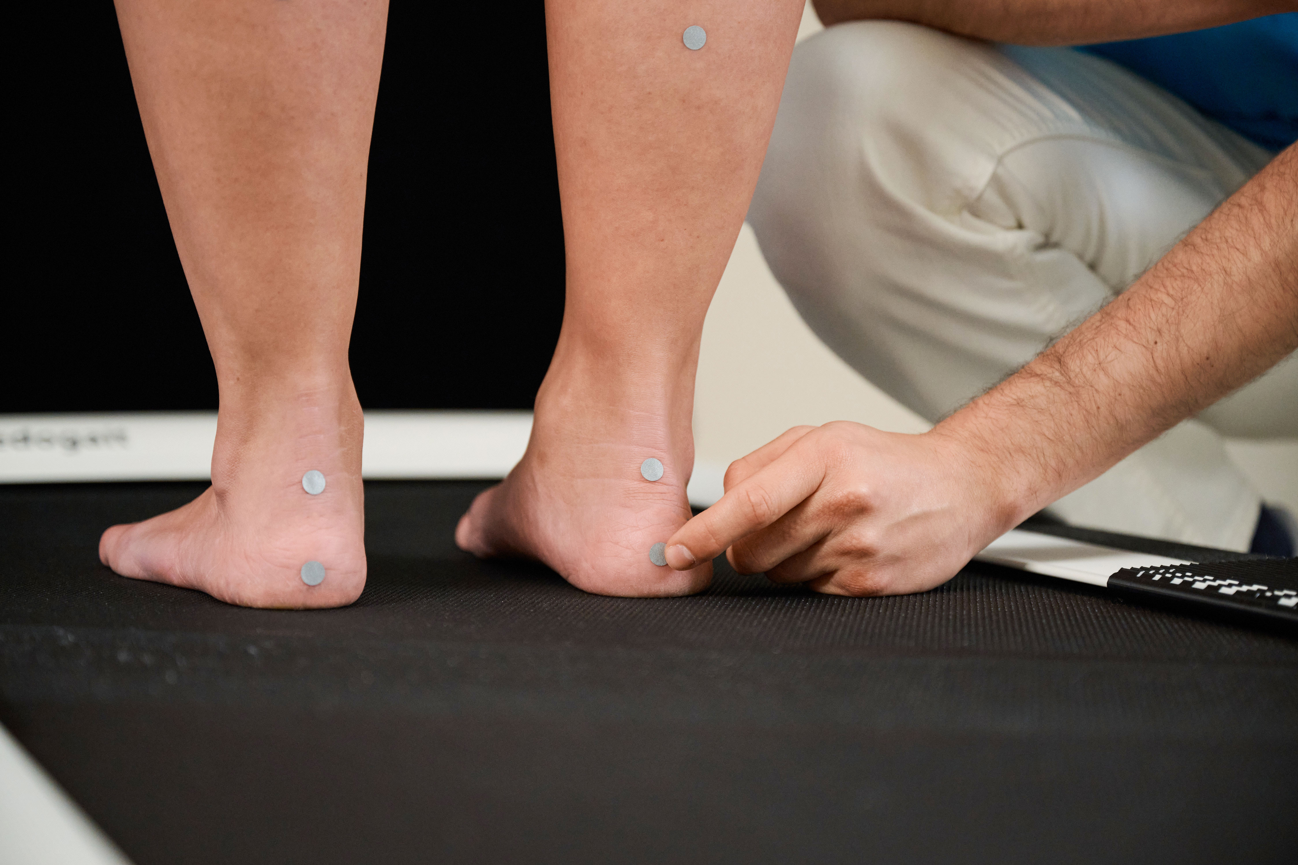
Precise analysis of pressure on your feet: we identify malalignment and develop targeted treatment to improve mobility and reduce symptoms.
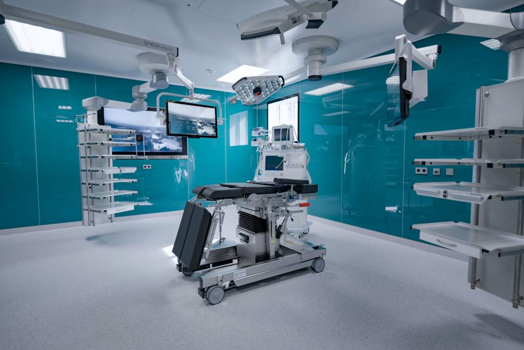
Modern techniques allow many procedures to be performed without an inpatient stay, safely, efficiently and tailored to the individual.
Case numbers, awards, quality reports and patient satisfaction show the standards against which we measure ourselves.