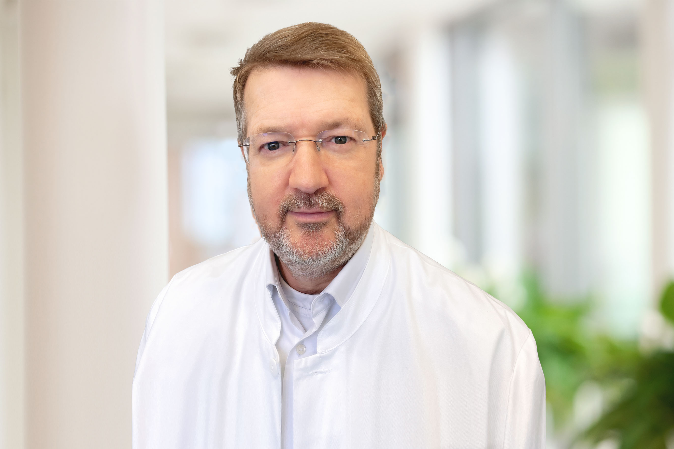
Dr med. dent. Manfred Suhr
Specialist in oral and maxillofacial surgery; dental specialist in oral surgery
Contact Dr med. dent. Manfred Suhr.
Jaw surgery, facial surgery, imaging and second opinions

Specialist in oral and maxillofacial surgery; dental specialist in oral surgery
Contact Dr med. dent. Manfred Suhr.
Skin conditions caused by ultraviolet light become more common with age. Because the face is exposed to particularly high levels of sunlight, it is often affected.
Alongside premature skin ageing, typical consequences of UV exposure include skin tumours. These range from precancerous changes and naevi, meaning pigmented marks or moles, to squamous cell carcinoma, basal cell carcinoma and malignant melanoma.
Simple pigmented marks such as naevi or moles are generally harmless. However, any change in colour, size or pigmentation should always be assessed histologically.
In addition to safely removing the skin lesion, particular attention is paid to functional and aesthetic reconstruction of important facial structures such as the eyelids, eyebrows, nose, ears and lips.
To achieve the least conspicuous scarring possible while preserving good function, incisions follow the relaxed skin tension lines and the aesthetic units of the face.
Skin conditions are always treated in close consultation and cooperation with the patient’s dermatologist.
The approach to malignant skin lesions is agreed by an interdisciplinary skin tumour board.
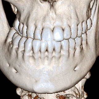
Jaw deformities have many causes, are often congenital and frequently cause functional as well as aesthetic impairment. Pathological changes in the jaw joints are also a common cause.
Healthy chewing and speech require the upper and lower teeth to meet correctly. Straightforward tooth misalignment can be corrected with orthodontic treatment using removable or fixed braces.
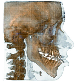
When the positions of the upper and lower jaw differ substantially, however, a functional bite can often be achieved only with surgery. This can frequently also create a more harmonious facial profile.
An individual treatment plan is developed in close consultation with the treating orthodontist using state-of-the-art digital 3D planning.
A visibly unusual nasal shape can have many different causes. It is often congenital and usually simply a normal variation; occasionally it is the result of an accident, such as a traumatic crooked nose. However, it is also frequently associated with substantial impairment of nasal function.
Misalignment of the upper jaw and midface, such as maxillary hypoplasia or midface hypoplasia, can also adversely affect the shape, function and projection of the nose.
This is often accompanied by narrowing at the entrance to the nose, known as the nasal valve, which can also obstruct nasal breathing.
An assessment by an ear, nose and throat specialist should always be carried out before planned functional and aesthetic nasal surgery.
Malformations of the outer ear, such as prominent ears, can be aesthetically distressing and can be corrected surgically using cartilage-preserving techniques.
A variety of causes can lead to loss of bone in the jaw. Tooth loss over a prolonged period commonly causes this type of jaw atrophy, which in turn makes artificial tooth replacement more difficult.
Modern medicines that have a positive effect on bone metabolism, such as bisphosphonates, are used very successfully to treat osteoporosis or bone metastases. However, medication-related osteonecrosis of the jaw is a recognised potential side effect.
A similar condition, known as osteoradionecrosis, can occur after radiotherapy for malignant tumours in the head and neck region.
Our aim is to reconstruct the affected parts of the jaw using the patient’s own bone or high-quality bone substitutes and, where needed, make high-quality dental restoration possible.
Conditions affecting the facial skeleton, oral mucosa and salivary glands are diagnosed according to the latest standards and treated appropriately.
Symptoms around the temporomandibular joint can have many different causes. The teeth, through the bite or occlusion, the chewing muscles and the two jaw joints form one functional unit whose balance is essential in this highly sensitive region. A disorder affecting any one of these structures can cause a complex pattern of symptoms throughout the functional unit. This is broadly described as temporomandibular disorder (TMD).
Clinical structural and functional analysis aims to identify the anatomical structure primarily affected and therefore the true source of pain. Grinding the teeth at night, for example, often causes muscle pain similar to soreness after exercise, which may lead to pain in the jaw joints months or years later. Conversely, painful rheumatic disease in a jaw joint can cause a painful reaction in the chewing muscles, creating a “chicken and egg” problem.
Uniquely in the human body, the two jaw joints can function only together. Pathological changes on one side therefore usually affect the other side as well. Another special feature is the jaw joint’s exceptionally wide range of movement, making it one of the body’s most mobile joints. This also makes it particularly susceptible to problems. Because the joint capsule is especially wide towards the front and centre, displacement of the articular disc in these directions is very common.
Very rare causes of symptoms in this area must also be ruled out during diagnosis, including tumours and congenital differences.
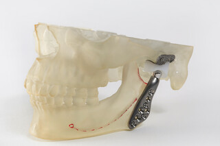
Even in advanced cases where the joint can no longer be preserved and complete replacement is required, modern patient-specific joint replacements provide a reliable treatment option. This may be considered, for example, where the joint has fused (ankylosis) or the condylar head is being resorbed.
Sleep-related breathing disorders can have serious health consequences and are often diagnosed only at a late stage.
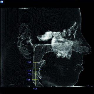
When the base of the tongue relaxes during sleep, a disorder can prevent enough air from passing through the throat, often causing snoring. The body does not receive enough oxygen. Patients typically report daytime tiredness and reduced performance. If left untreated, obstructive sleep apnoea syndrome (OSAS) often leads to chronic conditions such as high blood pressure and heart disease.
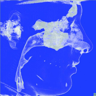
OSAS can also be caused by a receding lower jaw, which anatomically pushes the base of the tongue backwards.
A mandibular advancement device can be used to test whether moving the lower jaw forwards has a positive effect and, where appropriate, determine how much advancement is required.
Corrective osteotomy with advancement of the jaw, often involving the upper and lower jaw together, can then treat the underlying cause.
Case numbers, awards, quality reports and patient satisfaction show the standards against which we measure ourselves.